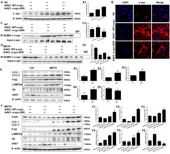
Figure 8.
Mutations in the major SUMOylation acceptor sites of α-syn exacerbate METH-induced α-syn aggregation in vivo. Adenovirus were injected into the right striatum of mice using a standard stereotaxic positioning system. After 2 days of recovery, mice received saline or METH by i.p. injection. Striatal tissues (right side) were harvested at 24 h after the injection of the last dose. The tissues were immunoprecipitated with an anti-SUMO-1 antibody, followed by western blot with an anti-α-syn antibody. Tissues immunoprecipitated with IgG were used as a negative control. Western blot (A,C) and quantitative analyses (A1,C1) were performed to evaluate the efficiency of WT α-syn and α-syn-KR expression. Immunofluorescence staining of mouse striatum sections showed that α-syn expression increased after injection of adenovirus (B). Western blot (D–F) and quantitative analyses (D1,E1–5,F1–6) were also performed to the levels of the SUMO-1, UbE1, Ub, LC3-II, P-62 and lysosomal associated membrane protein 2A (LAMP2A) proteins and the expression of SUMOylated α-syn. β-Actin was used as a loading control. *p < 0.05 compared with the AAV2-NC or control group. **p < 0.05 compared with the AAV2-α-syn group. #p < 0.05 compared with the AAV2-α-syn + METH-treated group. The data shown in (A,C,D,F) were analyzed using one-way ANOVA followed by LSD post hoc analyses, whereas the data shown in (E) were analyzed using the Mann-Whitney U test.

