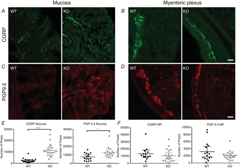
Figure 5. Neuronal sprouting in TNX‐KO mucosa.

Representative immunohistochemical images taken from sections of WT mouse colonic mucosa (A, C) and myenteric plexus (B, D). CGRP‐IR is shown in green (A) where there was a threefold proliferation of CGRP‐IR nerve endings in the colonic mucosa of TNX‐KO mice (P < 0.0001) (E). This was also observed with PGP9.5 in red (C), although this increase was smaller for PGP‐IR endings (P = 0.0224) (E). No changes were seen in either marker in the myenteric plexus (P = 0.3950, P = 0.1368) (F). Data are shown as individual values with standard deviation. Statistical analysis was performed using an unpaired Student's t test (P < 0.05). Scale bar in all panels = 20 μm.
