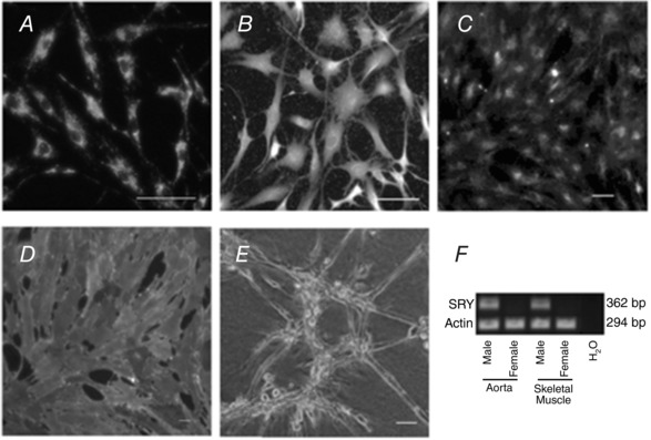
Figure 1. Verification of endothelial cell phenotype and sex.

Immunofluorescence staining of the endothelial cells was positive for PECAM‐1 in (A) and von Willebrand's factor in (B). The EC took up acetylated‐low density lipoprotein (Ac‐LDL) (C), stained positively with the lectin Griffonia simplicifolia IB4 (GS‐IB4) (D) and formed a tube‐like structure when plated on Matrigel (E). Scale bar = 50 μm. Male sex was confirmed using quantitative RT‐PCR for the presence of the sex‐determining region Y (SRY) protein (lanes 1 and 3) vs. its absence in endothelial cells from females (lanes 2 and 4). Actin was used as the loading control, whereas sterile water was used as a negative control (F).
