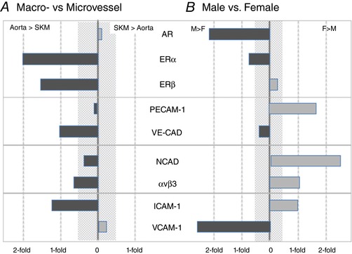
Figure 11. Ignoring sex (A) of tissue of origin (B) provides vastly differing data interpretations.

Overall assessment of the contributions of tissue origin (macro‐ vs. microvessel) in the absence of sex (A) and sex (male vs. female) in the absence of tissue origin (B) to protein expression. The shaded area represents differences of 50% or less, which is often difficult to detect reliably. For the proteins surveyed, overall expression levels appear to be higher in EC from the microvasculature and provide one image. In EC from males, androgen receptor (AR), ERα and VCAM levels are higher than females, ERβ and VE‐CAD display no sexual dimorphism, whereas PECAM‐1, NCAD, αvβ3 and ICAM‐1 levels are higher in EC from females relative to males. [Color figure can be viewed at http://wileyonlinelibrary.com]
