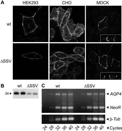
Figure 5.
AQP4 expression in cell lines transfected with wild-type (wt) AQP4 or AQP4 lacking three C-terminal residues (ΔSSV). (A) Transfected HEK293, CHO-K1, and MDCK cells labeled with anti-AQP4 and analyzed by immunofluorescence confocal microscopy show AQP4 wt and ΔSSV localized to the plasma membrane. Basolateral targeting in MDCK cells was confirmed in vertical sections (see Insets). (B) AQP4 immunoblot of membrane proteins prepared in duplicate from transfected HEK293 cells (20 μg protein per lane). (C) Expression of AQP4, neomycin resistance gene (NeoR), and β-tubulin (β-tub) assessed by RT-PCR of RNA from transfected HEK293 cells and ethidium bromide staining of agarose gels.

