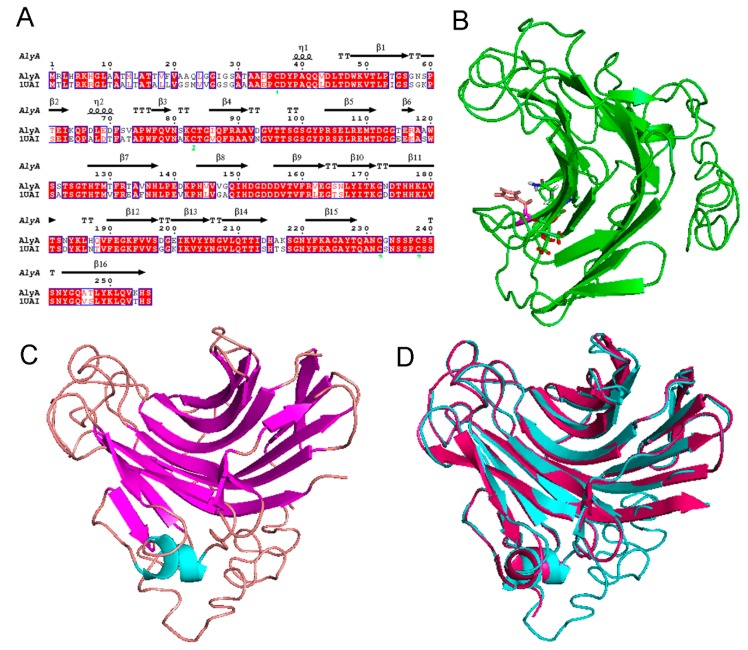
Figure 7.
(A) The sequence alignment of AlyA and AlyPG from Corynebacterium sp. ALY-1, (B) the modeling structure of AlyA, (C) the structural comparison of AlyA (marked with red) and AlyPG (marked with blue) from Corynebacterium sp. ALY-1 (PDB ID: 1UAI), and (D) the key residuals for substrate reorganization of AlyA.

