. 2018 Jul 3;32(4):745–752. doi: 10.21873/invivo.112303

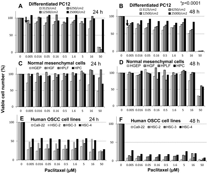
Figure 1. Cytotoxicity of paclitaxel towards rat neuronal cells compared to that towards human non-malignant and malignant cells. Differentiated rat PC12 cells (day 5) inoculated at different density (3,125-25,000 cells/cm2) (A, B), human normal epithelial (HGEP) and mesenchymal cells [human gingival fibroblast (HGF), human periodontal ligament fibroblast (HPLF), human pulp cell (HPC)] (C, D), and human oral squamous cell carcinoma cell lines derived from gingiva (Ca9-22) and tongue (HSC-2, HSC-3, HSC-4) (E, F) were treated for 24 (A, C, E) or 48 h (B, D, F) with the indicated concentrations of paclitaxel, and relative viable cell number was determined by MTT method. Each value represents the mean from six determinantions. The 50% cytotoxic concentration (CC50) of paclitaxel is listed in Table I.

Figure 1