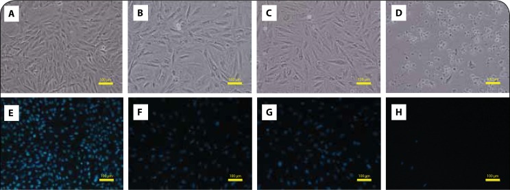
Figure 2.
PIX causes cytotoxicity in non-differentiated H9c2 without signs of condensed nuclei. Phase contrast microscopy (A, B, C, D) and fluorescence microscopy (Hoechst 33258 staining) (E, F, G, H) images of non-differentiated H9c2 cells after a 48-h exposure to PBS (A and E), 0.1 μM PIX (B and F), 1 μM PIX (C and G) or 10 μM PIX (D and H). Images are representative of two independent experiments (scale bar 100 μm).

