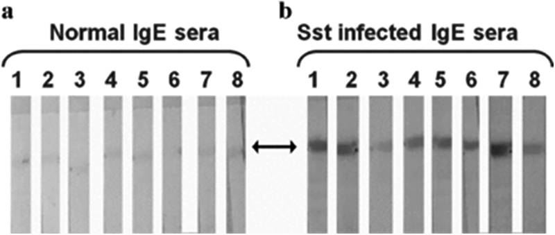
Figure 5.
Western blot analysis of IgE antibodies in sera from control and S. stercoralis infected patients probed with purified strongylastacin antigen. Purified strongylastacin antigen was separated on NuPAGE gel and strips were made after transferring protein from the gel to the nitrocellulose membrane. Both normal and S. stercoralis infected human sera were pre‐absorbed with protein G sepharose to enrich IgE antibodies. Strips were separately incubated with 1:8 times diluted sera for 1 hr. Blots were stained with a second antibody labeled with goat anti‐ human IgE antibody conjugated to alkaline phosphatase. Panel (a) none of the eight normal sera reacted. Panel (b) IgE antibodies in seven of eight strongly reacted with strongylastacin and one showed weak reactivity. Sst, S. stercoralis.

