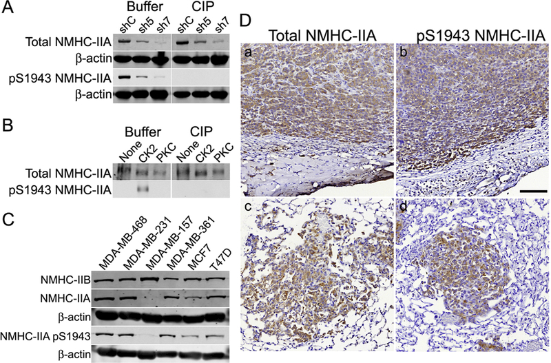
Fig. 1.

Characterization of pS1943 NMHC-IIA antibodies. (A) MDA-MB-231 cells were transduced with lentivirus expressing control (shC) or shRNAs (sh5 and sh7) against human NMHC-IIA. Buffer- or calf intestinal and lambda phosphatase (ClP)-treated immunoblots were blotted for total NMHC-IIA, pS1943 NMHC-IIA (Millipore antibody) or β-actin. (B) Buffer- or CIP-treated immunoblots (total and pS1943 NMHC-IIA - Millipore antibody) of unphosphorylated, S1943-phos- phorylated (CK2) and S1916-phosphorylated (PKC) myosin-IIA rods. (C) Representative immunoblots showing expression of total NMHC-IIA, NMHC-IIB, pS1943 NMHC-IIA (Millipore antibody) or β-actin in a panel of breast cancer cell lines. (D) Immunohistochemistry of MDA-MB-231–3475 orthotopic tumors (a, b) and spontaneous lung metastatic nodules (c, d), showing expression of total NMHC-IIA or pS1943 NMHC-IIA (Cell Signaling Technology antibody), which appears as a brown stain. Scale bar = 100 μm.
