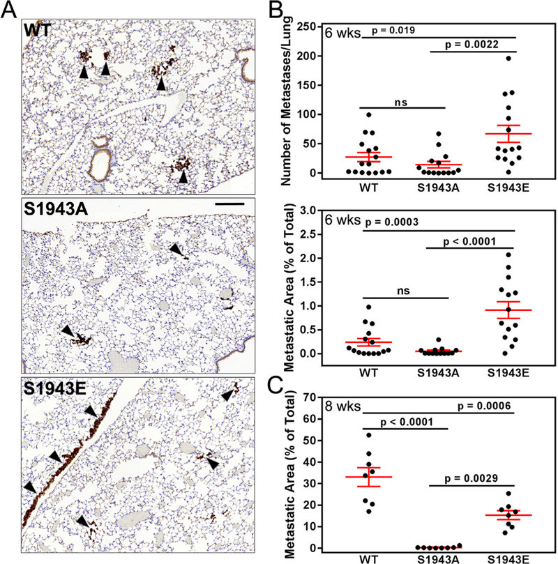
Fig. 7.

S1943 NMHC-IIA phosphorylation regulates extravasation in vivo. (A) Pan-cytoker- atin stained lung sections from SCID mice injected with MDA-MB-231 cells expressing wild- type, S1943A or S1943E NMHC-IIA six weeks after injection of tumor cells. Arrowheads indicate metastatic foci. Bar = 200 μm. (B) The number and percent area of metastases per lung section six weeks after injection of tumor cells. Data represent the mean ± SEM from two independent experiments with a total of 15, 13, and 14 mice for wild-type, SI943A, or S1943E NMHC-IIA cells, respectively. Statistical analyses were performed using ANOVA. (C) The percent area of metastases per lung section eight weeks after injection of tumor cells. Data represent the mean ± SEM from 8 mice each for wild-type, S1943A or S1943E NMHC-IIA cells, respectively. Statistical analyses were performed using ANOVA.
