
Figure 2.
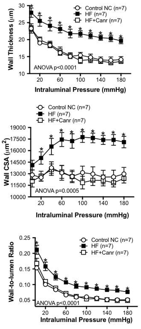
MR is involved in the hypertrophic growth of the MCA wall in obese rats. Chronic HF treatment caused a dramatic hypertrophy of the MCA wall, observed as an increase in the wall thickness (A) and the wall CSA (B). The wall hypertrophy was prevented in HF rats that received the MR antagonist Canr. As consequence of the increase in wall thickness, the wall-to-lumen ratio of MCAs from rats fed HF alone was higher than rats fed Control NC or HF+Canr (C), suggesting an inward hypertrophic remodeling. MCAs were bathed in warm (37°C), oxygenated (95% O2) Ca2+-free PSS supplemented with 2mmol/L EGTA and 10μmol/L SNP. MCAs were allowed to equilibrate for 5 minutes at each intraluminal pressure before measurement was performed. *p<0.001, statistically different from Control NC; αp<0.001, statistically different from HF+Canr, two-way ANOVA with a Tukey’s post-test for multiple comparisons.