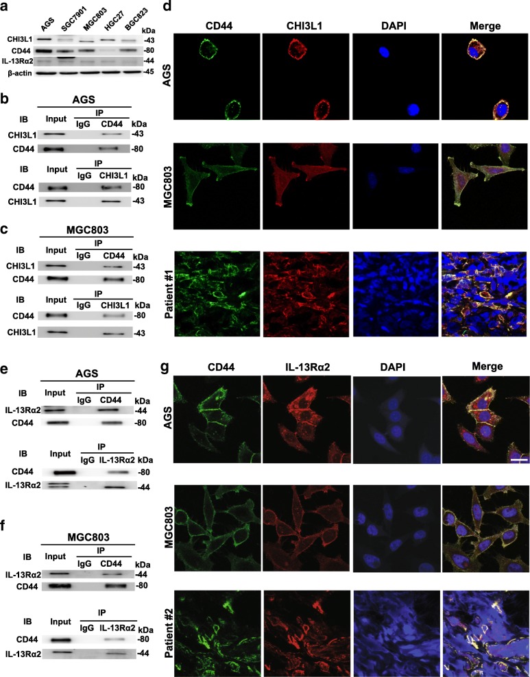
Fig. 2.
CHI3L1 binds to CD44 which also interacts with IL-13Rα2. a Western blot analysis of CHI3L1, CD44 and IL-13Rα2 protein expression in various gastric cancer cell lines. b-c Lysates from AGS and MGC803 cells were immunoprecipitated (IP) with control IgG and anti-CD44 or anti-CHI3L1 antibody, and then immunoblotted as indicated. Five percent of total cell lysates were used for the input. d Co-localization of CD44 (green) and CHI3L1 (red) in AGS (upper panel), MGC803 (middle panel) and GC tissues from patient #1 (lower panel) by immunofluorescent confocal microscopy (Magnification: 630×). Scale bars represent 10 μm. e-f Lysates from AGS and MGC803 cells were immunoprecipitated (IP) with IgG and anti-CD44 or anti-IL-13Rα2 antibody, and then immunoblotted as indicated. Five percent of total cell lysates were used for the input. g Co-localization of CD44 (green) and IL-13Rα2 (red) in AGS (upper panel), MGC803 (middle panel) and GC tissues from patient #2 (lower panel) by immunofluorescent confocal microscopy (Magnification: 630×). Scale bars represent 10 μm

