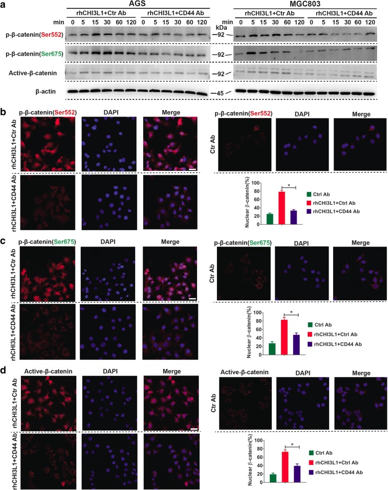
Fig. 4.
CHI3L1 regulates β-Catenin signaling via CD44. a Western blot analyses were used to evaluate β-catenin phosphorylation (Ser675 or Ser552), and active-β-Catenin (ABC) after exposure to rhCHI3L1 (500 ng/ml) in the presence of control or CD44 neutralizing antibody (10 μg/ml) for the noted periods of time. b-d AGS cell were pretreated with control IgG or CD44 blocking antibody (10 μg/ml). Then, the immunofluorescence assays of p-β-catenin (Ser552 or Ser675) and active-β-catenin (ABC) (all in red) were performed in AGS cell treated with rhCHI3L1 (500 ng/ml). DAPI (blue) was used as a nuclear counterstain. The quantification of nuclear β-catenin positive staining in at least 200 counted cells was presented as percentage ± SEM. Magnification: 400×, Scale bars represent 20 μm. Results shown here are the representative of three independent experiments. Statistical significance was calculated using ANOVA (b-d). *p < 0.05

