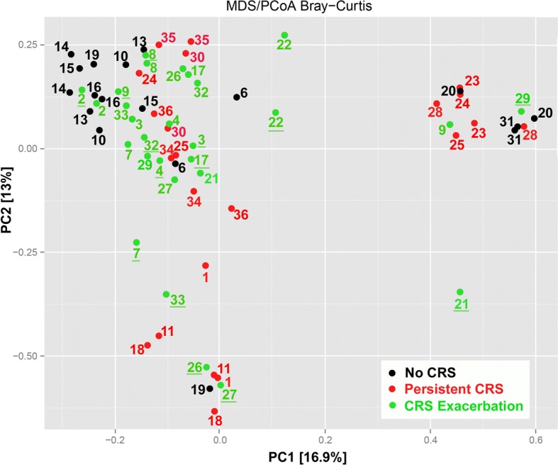
Fig. 5.

Principal coordinate analysis of Bray–Curtis dissimilarity index generated from taxa of the three groups at both time points. Proportion of variance explained by each principal coordinate axis is denoted in the corresponding axis label. Colored by group and tested with ANOSIM (R = 0.28, p = 0.001). Black circles: group 1; red circles: group 2; green circles: group 3. Inflamed time points for group 3 are underlined
