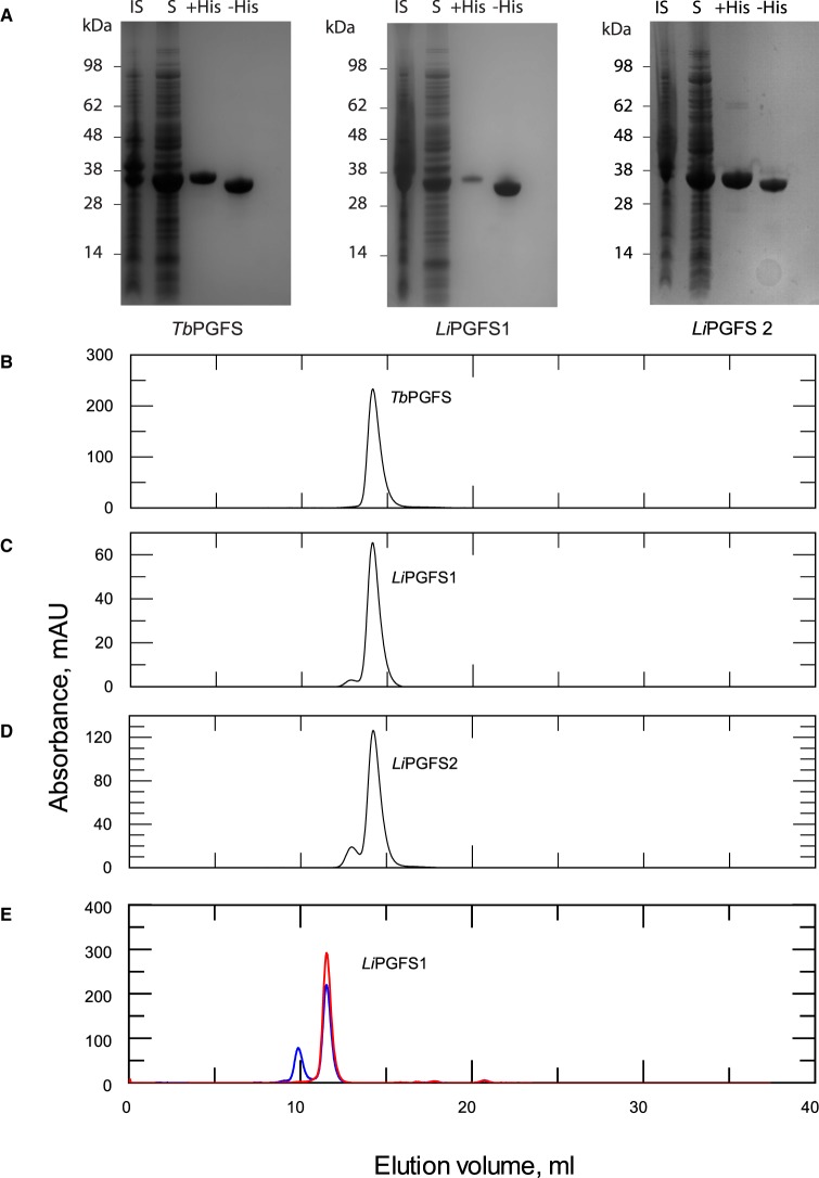
Figure 5. Characterisation of LiPGFS1, LiPGFS2 and TbPGFS.
(A) SDS–PAGE showing purification of TbPGFS1, LiPGFS1 and LiPGFS2 showing insoluble, soluble, pooled protein from HisTrap column and protein with the hexahistidine tag removed. Size exclusion elution profiles of (B) TbPGFS, (C) LiPGFS1 and (D) LiPGFS2. Samples were separated on a Superose 12 10/300 column in 25 mM HEPES, 150 mM NaCl, pH 7.33. (E) LiPGFS1 stored in the absence of a reducing agent analysed by size exclusion chromatography in the absence (blue) and presence (red) of 0.5 mM TCEP using a Superdex 75 10/300 column.

