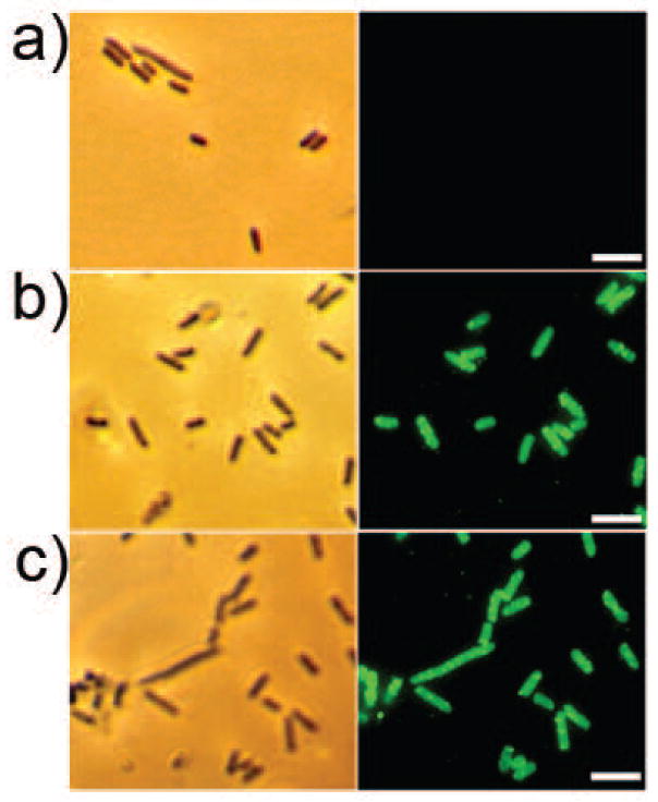
Figure 2.
Immunofluorescence of Rosetta (DE3) pLysS cells. a) Control Rosetta (DE3) pLysS cells transformed with pET29b. b) Rosetta (DE3) pLysS cells mobilized with pKD11 encoding the T7 tag. c) Rosetta cells with pKD3 encoding dispersin B. Shown on the left are the phase contrast images and on the right, immunofluorescence images. The bar indicates 5 μm. Green fluorescence was due to Alexa Fluor 488 labeled secondary antibody.

