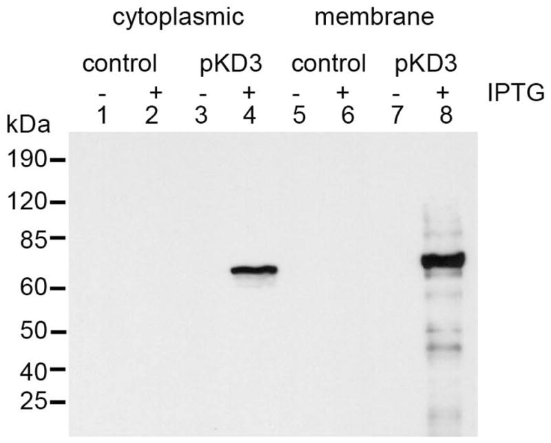
Figure 4.

Western blot analysis of DspB surface localization. The cytoplasmic as well as membrane fractions were boiled in sample buffer. The samples were run on 10% SDS-PAGE, transferred to a membrane and probed using polyclonal anti-DspB antibody. Lanes 1, 2, 5 and 6 were from control Rosetta (DE3) pLysS cells. Lanes 3, 4, 7 and 8, are from the pKD3 cells. Odd numbered lanes were not induced with IPTG (0.1 mM) whereas even numbered lanes were induced with IPTG.
