Figure 1.
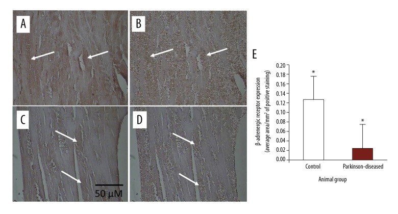
Immunohistochemical staining of β-adrenergic receptor in longitudinal 4-μm-thick paraffin-embedded heart sections. (A) From control. (C) From Parkinson-diseased (PD). Scale bar shown in (C) applies to all images in the figure. β-adrenergic receptor immunostaining was very strongly observed in the control hearts. In contrast, β-adrenergic receptor immunoreactivity is weak in hearts from the PD group (such as those at the tips of the arrows). Areas of positive immunostaining of β-adrenergic receptor, shown in brown at the tips of the arrows in control (A) and PD (C) heart sections, are mapped as pixel areas at the tips of the arrows (B, D, respectively). (E) β-adrenergic receptor levels decreased significantly in the PD hearts compared to those in the control group (* P<0.01).

