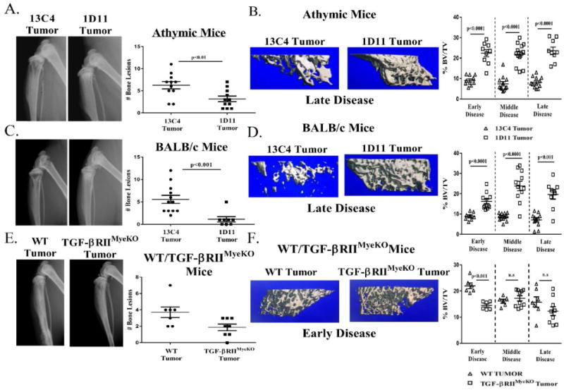
Figure 6. TGF-β inhibition reduces osteolytic lesions and improves overall bone volume.

X-ray analysis of tumor-bearing mice at late stage disease prior to sacrifice. Representative images are from late stage disease. Formalin fixed tibias were scanned and analyzed by μCT as described in the methods section. Percent bone volume normalized to total volume (BV/TV) for each treatment group. A. Athymic mouse model: X-ray images, 13C4 treated (left), 1D11 treated (right) and quantification of bone lesions. Tumor-bearing athymic mice treated with 1D11 have less osteolytic lesions than isotype control. B. Left: three-dimensional (3D) rendered μCT images from two tumor-bearing athymic mice at late stage disease, 13C4 treated (left), 1D11 treated (right). Right: percent BV/TV over the course of tumor-induced bone disease (TIBD) in the athymic mouse model. Treatment with 1D11 increases overall bone volume over the course of TIBD. C. BALB/c mouse model: X-ray images, 13C4 treated (left), 1D11 treated (right) and quantification of bone lesions. Tumor-bearing BALB/c mice treated with 1D11 have less osteolytic lesions than isotype control. D. Left: 3D rendered μCT images from two tumor-bearing BALB/c mice at late stage disease, 13C4 treated (left), 1D11 treated (right). Right: percent BV/TV over the course of tumor-induced bone disease in the BALB/c mouse model. Treatment with 1D11 increases overall bone volume over the course of TIBD. E. WT/TGF-βRIIMyeKO mice mouse model: X-ray images, WT tumor (left), TGF-βRIIMyeKO tumor (right) and quantification of bone erosions. Tumor-bearing TGF-βRIIMyeKO have a reduced incidence of bone lesions as compared to tumor-bearing WT mice. F. Left: 3D rendered μCT images from two tumor-bearinF-βRIIMyeKO (right). Right: percent BV/TV over the course of tumor-induced bone disease in the WT/TGF-βRIIMyeKO mice mouse model. Tumor-bearing TGF-βRIIMyeKO mice only have a significant decrease in their overall bone volume at early disease. (9-13 tibias per group).
