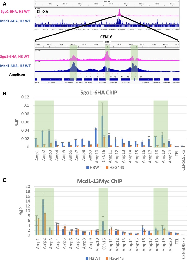
Figure 3.
The histone H3 tension sensing motif is essential for pericentric Sgo1p localization but not Mcd1p. A. Distribution of Sgo1p (magenta) and Mcd1p (blue) across chromosome XVI as revealed by ChIP-seq. The centromeric region is blown up to show the detail distribution of these two proteins. PCR amplicons are enumerated and shown in light pink bars below the Mcd1 peaks. The open reading frames and their transcription directions are shown at the bottom. B and C. Quantitative real-time PCR analysis of separate ChIP experiments. Sgo1p-HA and Mcd1p-Myc (both expressed from their native loci) were ChIP’ed from cells bearing the wildtype or a mutant TSM (G44S). The three enrichment sites are marked with shaded boxes. ChIP-qPCR data were from three biological replicas. We repetitively observed that the Mcd1p ChIP signals to be significantly higher than those of Sgo1p (also see Figure 5). This differentiation may result from the choice of the epitope tags (13-Myc vs. 6- or 3-HA), or the nature of chromosome association, or both.

