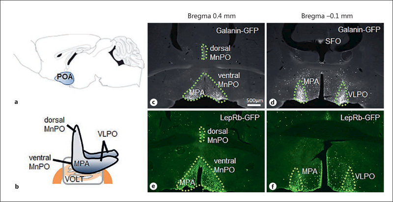
Fig. 1.
a Sagittal scheme of the mouse brain highlighting the gross location of the preoptic area (POA) within the CNS. b–f The POA is associated with the vascular organ of the lamina terminalis (VOLT) and the subfornical organ (SFO), both circumventricular organs that allow access and exchange with the circulation. Furthermore, several subareas can be distinguished in the POA that are well visualized by galanin- or leptin receptor (LepRb)-expressing neurons: dorsal and ventral median preoptic area (MnPO), medial preoptic area (MPA), and the ventrolateral preoptic area (VLPO).

