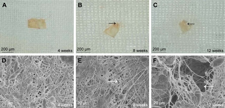
Figure 8.
The morphology of the ICA-loaded PCL–gelatin membrane (A–C), and SEM micrographs of membrane surfaces (D–F) at different observation times after implantation.
Note: The black arrows indicate the absorbed margin, and the white arrows indicate the pores.
Abbreviations: ICA, icariin; PCL, polycaprolactone; SEM, scanning electron microscopy.

