
Figure 1. Photomicrographs of histological and immunohistochemical studies of representative gastric and surgical lung biopsy specimens.
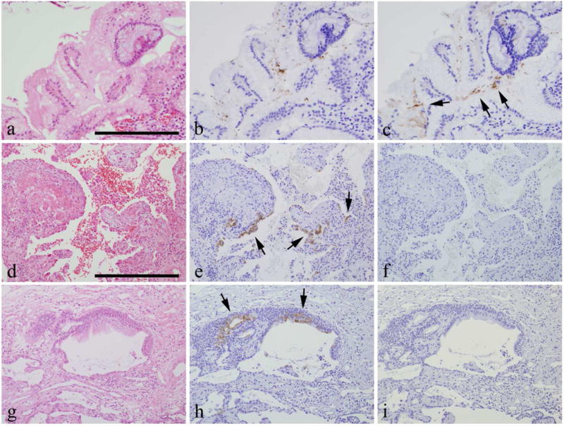
a–c are sequential sections, a: Hematoxylin-eosin staining, b: Immunohistochemistry with anti-Helicobacter pylori antibody, c: Immunohistochemistry with anti-VacA antibody. Arrows show VacA. Scale bar: = 200 μm.
d–f and g–i are sequential sections obtained from 2 patients. Lung tissues in d–f and gastric tissues in a–c were obtained from the same patient, d, g: hematoxylin-eosin staining, e, h: immunohistochemistry with anti-VacA antibody. Arrows show VacA. f, i: control studies using rabbit immunoglobulin as the primary antibodies. Scale bar: = 400 μm.