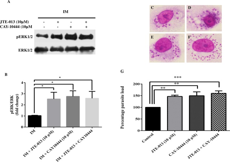
Fig 6. Anti-inflammatory effect on inhibition of S1PR2-3 in infected macrophages.
TDM were cultured in six-well plates in the presence or absence of L. donovani infection (MOI = 1: 10) for 6 h, TDM were washed to remove non-internalized parasites and incubated for next 42 h in presence and absence of JTE-013 and CAY-10444. A. Western Blot showing increase in the phosphorylation of ERK1/2 in presence of S1PR2 inhibitor (JTE-103) or S1PR3 inhibitor (CAY10444) or both in IM. B. Densitometric analysis of ERK1/2 in presence of JTE-013 and CAY10444 in infected and uninfected macrophages, after normalization with total ERK1/2. Giemsa stained L. donovani infected TDM. C. Control, D. JTE-013(10 μM) treated, E. CAY-10444 (10 μM) treated, F. JTE-013 (10 μM) and CAY10444 (10 μM) treated. G. The % parasite load was determined by direct counting with an optical microscope. The data is a representation of mean ± SD from three independent experiments. * p < 0.05, ** p < 0.01, ***, p < 0.001.

