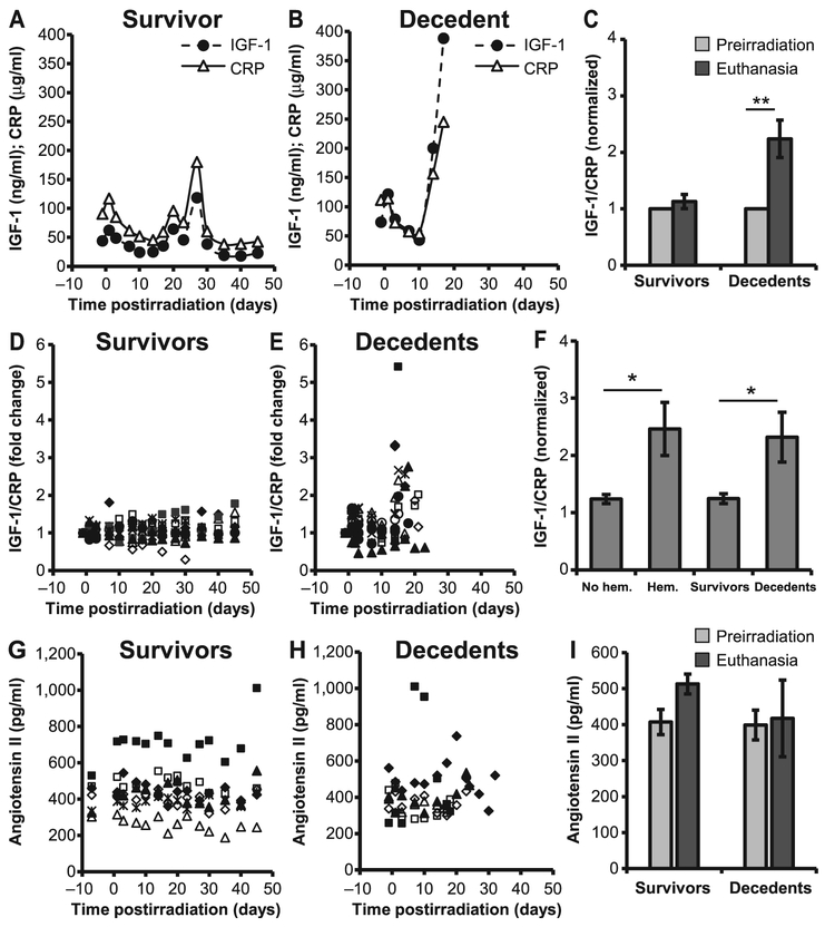
FIG. 3.
Dynamic changes in the expression of IGF-1 and CRP. Plasma IGF-1 and CRP levels were compared longitudinally in survivors and decedents (panels A, B, D and E). Representative IGF-1 and CRP levels from one survivor and one decedent (panels A and B, respectively) show that the expression of IGF-1 paralleled that of CRP, however, the equilibrium was lost when the animals became sick due to radiation injury (panel B). Fold changes in the IGF-1-to-CRP ratio were higher at euthanasia than preirradiation in decedents (panel C). IGF-1 and CRP expression levels from individual animals were normalized to their preirradiation levels and are shown as the relative fold changes for survivors (panel D) and decedents (panel E). Panel F: The ratio of CRP to IGF-1 in animals with and without hemorrhages (hem) in heart and also decedents compared to survivors. Values plotted in panels C and F are mean average ratios after normalization. Panels G–I: Plasma angiotensin II concentration from individual survivors (panel G), decedents (panel H) and average angiotensin II levels at preirradiation and at euthanasia (panel I). There were no statistically significant differences in the concentration of angiotensin II in decedents compared to survivors. Values plotted in panels C, F and I are mean ± SEM. Data analyzed using Student’s paired t test; n = 10–12/group. *P < 0.05; **P < 0.01. Each symbol in panels D, E, G and H represent an individual animal.

