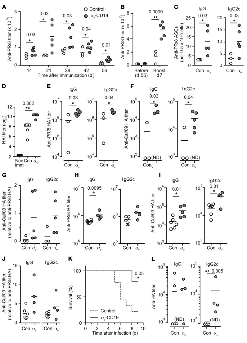
Figure 9. Deletion of αv enhances antibody response to influenza virus.
(A and B) Serum anti-PR/8 IgG titers in control and αv-CD19 mice immunized with 10 μg of inactivated H1N1 PR/8 (A) and (B) boosted at day 57 with 5 μg inactivated PR/8. (C) PR/8-specific plasma cells enumerated by ELISpot in BM cells from control (Con) and αv-CD19 mice harvested at day 7 after boost. (D) HAI activity in sera from control and αv-CD19 mice at day 21 after PR/8 immunization. (E and F) Serum antibody titers against HA from H1N1 PR/8 (E) or H1N1 Cal/09 (F) in control and αv-CD19 mice at day 7 after boost with inactivated PR/8. (G) Anti-Cal/09 HA titer normalized to anti-PR/8 HA titer. (H and I) Serum antibody titers against HA from PR/8 (H) or Cal/09 (I) in control and αv-CD19 at day 51 after immunization with inactivated PR/8 (10 μg) in imiquimod-SE (10 μg). (J) Anti-Cal/09 HA titer normalized to anti-PR/8 HA titer for mice immunized with PR/8 in imiquimod-SE. (K) Survival of control and αv-CD19 mice following intranasal infection with PR/8 (n ≥ 5 mice/group). (L) Anti-PR8 HA titers from surviving mice at day 7 after infection. All data points represent individual mice with mean shown. P values of less than 0.05 are shown (Mann-Whitney-Wilcoxon test for antibody titers or Mantel-Cox test for survival curves).*P < 0.05; **P < 0.005. Samples below the level of detection are indicated as not detected. For all data, similar results were seen in at least 3 independent experiments.

