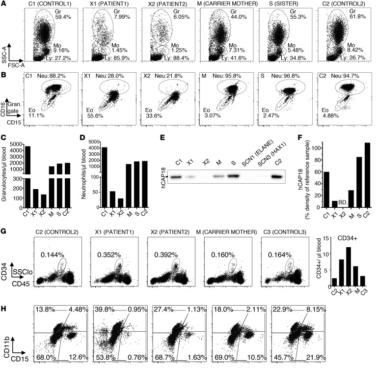
Figure 1. Severe neutropenia with hyperactivated neutrophils in the blood of XLN patients.
(A) Forward and side scatter flow cytometry plots of RBC-lysed blood from WASp L270P XLN patients (X1 and X2), their mutation carrier mother (M), their sister with unknown carrier status (S), and 2 healthy male controls (C1 and C2). Granulocyte (Gr), monocyte (Mo), and lymphocyte (Ly) gates are marked with circles. (B) CD15 vs. CD16 staining of whole-blood granulocytes. Eo, eosinophils; Neu, neutrophils. (C and D) Granulocyte and neutrophil numbers in blood counted (from A and B populations) with flow cytometry using counting beads. (E) hCAP18 expression in serum as determined by Western blot. Serum from SCN1 (ELANE) and SCN3 (HAX1) patients served as references. (F) Densitometry of hCAP18 blots indicated as percentage density of 1 μg/ml hCAP18 reference sample. BD, below detection limit. (G) Percentage and number of CD34+ hematopoietic progenitors in the blood of C2, X1, X2, M, and C3 (female control). (H) Composition of in vitro–differentiated myeloid liquid cultures from CD34+ blood cells.

