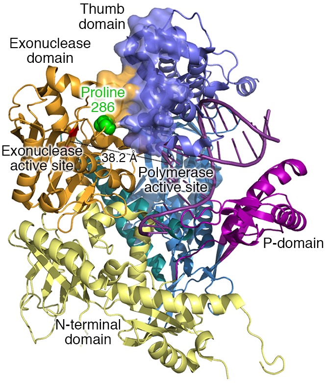
Figure 1. X-ray crystal structure of DNA polymerase ε.

X-ray crystal structure of the 142-kDa N-terminal region of the catalytic subunit of yeast DNA polymerase ε (10). Yeast proline 301 is homologous to human proline 286, which is highlighted in green. The catalytic residues for the 3′ exonuclease activity are shown in red. They are located 38.2 Å away from the polymerase active site. See text for further descriptions.
