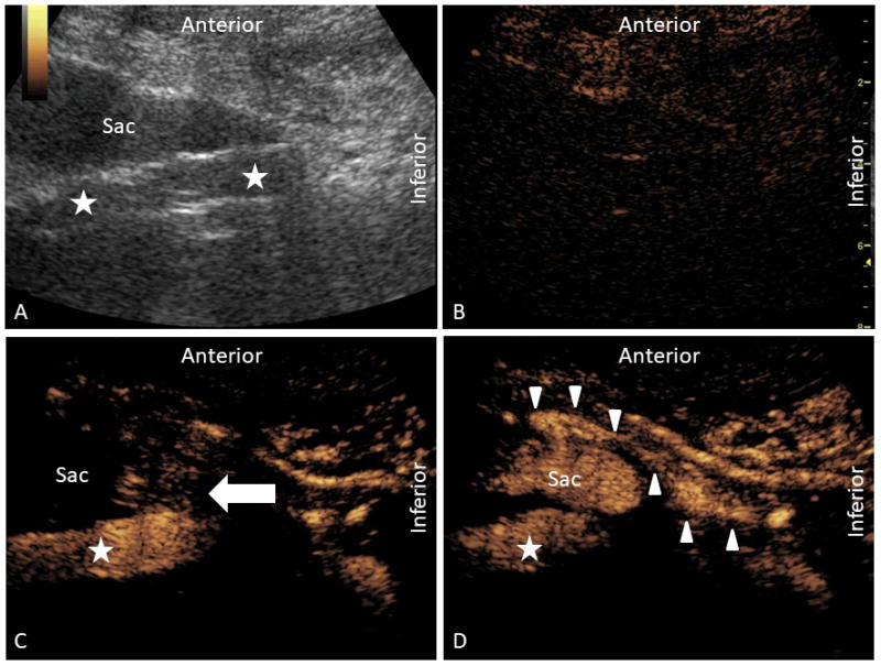
Figure 2.

Contrast-enhanced ultrasound examination depicting a complex multi-type endoleak in the sagittal orientation. In panel A, grayscale ultrasound of the aorta (star) is depicted with an anteriorly positioned aneurysmal sac. In panel B, contrast-mode is depicted prior to the administration of contrast. At the current gain setting, the aorta is just out of view. In panel C, a type III endoleak is demonstrated (arrow) at the junction of the body and left iliac extension limb. In panel D, a type II endoleak is demonstrated by reversal of flow within the traced inferior mesenteric artery (arrowhead) and perfusion into the sac.
