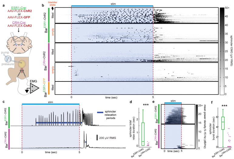
Fig. 5. BarESR1 neurons relax the urethral sphincter by promoting bursting activity.
a, Schematic for optogenetic Bar stimulation during cystometry and urethral EMG recording. b, Heatmap of EUS EMG, sorted by increasing mean voltage, around photostimulation for both filled and empty bladder trials (n=6 BarESR1-ChR2, top, 5 BarCRH-ChR2, middle, and 3 BarESR1-GFP, bottom, mice). c, Example RMS EMG traces from single ‘filled’ bladder trials in (b), showing calculated sphincter relaxation periods (dark blue) between bursts. d, Total sphincter relaxation time boxplot (median, 25th/75th quartiles, 1.5x interquartile ranges, and outlier dots outside these ranges) for ‘filled’ bladder trials in (b), n=33 BarESR1-ChR2 trials and n=48 BarCRH-ChR2 trials, ***p=2.4E-8 (Mann-Whitney U test). e, Heatmap of mean EMG power density at bursting frequencies (5–15 Hz) for ‘filled’ bladder trials in (b) (BarESR1-ChR2, top, and BarCRH-ChR2, bottom). f, Sphincter burst duration boxplot for trials in (e), n=33 BarESR1-ChR2 trials and n=48 BarCRH-ChR2 trials, ***p=1.1E-11 (Mann-Whitney U test). Quartile range boxes in (d) and (f) are condensed at zero when most trials do not have any bursting.

