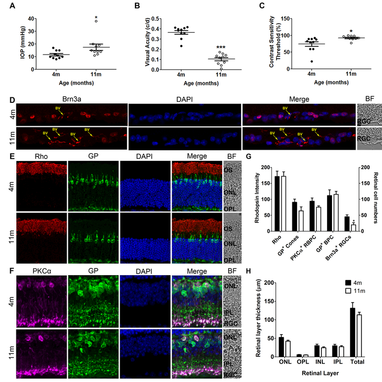
Figure 1:
Eleven-month old DBA/2 mice have increased IOP (A), reduced visual function [measured as reduced visual acuity (B) and increased contrast sensitivity threshold (C)] and significant RGC loss (Brn3a-positive cells in D [red label], G) indicative of glaucoma disease progression and when compared to young, non-glaucomatous control mice. Photoreceptor (E, G) and bipolar cell numbers (F, G) and retinal layer thickness (H) in 11-month old glaucomatous mice are not significantly different from 4 month-old non-glaucomatous mice; data are expressed as the number of cells per 1-mm retinal length in a vertical section or the thickness of the retina or layer in µm. In E and F, rhodopsin (Rho) immunoreactivity was found in the outer segments of the rod photoreceptors (red); immunoreactivity for Glycogen Phosphorylase (GP) labelled cone photoreceptors, cone bipolar cells and their terminals (green), and for Protein Kinase α (PKCα) labelled rod bipolar cells and their terminals (purple) for Brn3a labelled RGCs. Nuclei were stained with DAPI (blue). BV= Blood vessel. Data are expressed as mean ± SEM.

