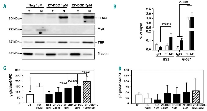
Figure 1.
Increased γ-globin gene transcription in sickle erythroid progenitors after delivery of the -567GγZF-DBD. Peripheral blood mononuclear cells isolated from sickle cell patients were used to induce differentiation along the erythroid lineage according to a previously published method.5 On day 8 of differentiation (see Online Supplementary Methods), cells were exposed for 48 hours (h) to HU, NC-ZF-DBD (Neg), or -567GγZF-DBD (ZF-DBD) at the concentrations shown. Experimental conditions were tested in triplicate for the 3 sickle cell patients (n=9) for each completed data analysis. (A) Localization of NC-ZF-DBD and ZF-DBD in the nucleus after protein delivery. Proteins were isolated from the cytoplasm (C) or nucleus (N) as previously published;4 western blot analysis was conducted as described previously15 using antibodies specific for the -567GγZF-DBD (aFLAG, F3165, Sigma, St. Louis, MO, USA), the NC-ZF-DBD (aMyc, MA121316, Thermo Fisher Scientific, Waltham, MA, USA), nuclear TATA-Binding Protein (aTBP, SC-204, Santa Cruz Biotechnology, Dallas, TX, USA) and b-actin (b-actin, A5316, Sigma, St. Louis, MO, USA). (B) Specific interaction of the -567GγZF-DBD with the Gγ-globin upstream promoter region. Cells were exposed to 1 µM (white bars) or 3 µM ZF-DBD (black bars), crosslinked with formaldehyde, and subjected to ChIP assay as described previously15 using -567GγZF-DBD (aFLAG) or IgG control (I8140, Sigma, St. Louis, MO, USA) antibodies. The purified DNA was analyzed by quantitative (q) PCR using primers specific for LCR HS2 and the Gγ-globin upstream promoter (G-567). (C and D) To measure gene transcription rates, reverse transcriptase-qPCR was conducted using RNA isolated from sickle erythroid progenitors after 48 h of treatment (day 10) in culture (see Online Supplementary Methods). Increased γ-globin (C) but not bS-globin (D) gene transcription was observed in cells exposed to hydroxyurea (HU), the -567GγZF-DBD (ZF-DBD), or both but not in cells treated with the NC-ZF-DBD (Neg) alone. RNA was extracted from sickle erythroid progenitors treated under the different conditions shown, reverse transcribed, and subjected to qPCR using primers specific for GAPDH, γ-globin, and bS-globin as described previously.15 The results in (B), (C), and (D) are from 3 independent experiments and error bars reflect the Standard Deviation (SD).

