Fig. 3.
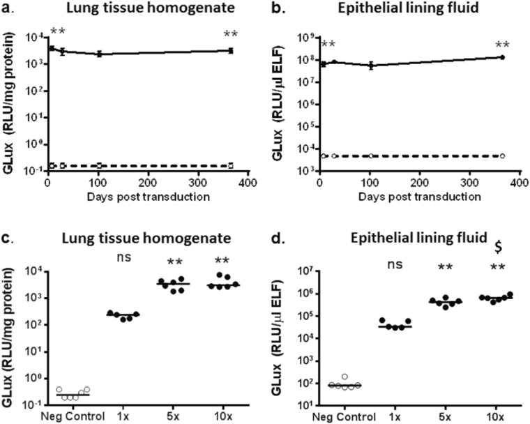
Sustained and dose-related expression of Gaussia luciferase (GLux) in mice after lentivirus transduction. C57Bl/6 mice were transduced with rSIV.F/HN carrying the soGLux cDNA under the control of the hCEF promoter (rSIV.F/HN-hCEF-soGLux, 1e7 TU/mouse, solid line) or received D-PBS (negative control, dotted line) by nasal sniffing and were culled between 7 and 365 days post transduction. GLux expression was measured in (a) lung tissue homogenate and b epithelial lining fluid (ELF). Data are expressed as mean ± SEM, n = 5–6 per group. ** = p < 0.01 compared with the negative control (only the early and late time-point were compared statistically using analysis of variance followed by a Bonferroni post hoc test. In a separate experiment using a different batch of virus mice were transduced with one, five or 10 doses of rSIV.F/HN-hCEF-soGLux and culled 7 days after the last dose. Owing to technical reasons the titre of the batch of virus could not be determined, but this is unlikely to affect the interpretation of the data. Negative control animals were treated with 10 doses of D-PBS. GLux expression was measured in (c) lung tissue homogenate and d ELF. Each dot represents one animal and the horizontal line represents the group mean. Data are expressed as mean ± SEM, n = 5–6 per group. RLU = relative light units. ** = p < 0.01 compared with negative control. $ = p < 0.05 compared with 5 × ns = not significant following correction for multiple comparison using analysis of variance followed by a Bonferroni post hoc test

