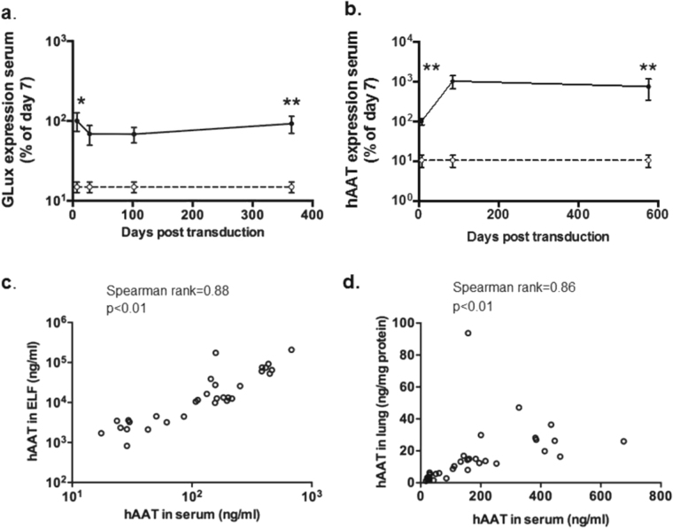
Fig. 5.
Release of recombinant proteins from lung into the circulation. a C57Bl/6 mice were transduced with rSIV.F/HN-expressing Gaussia luciferase (rSIV.F/HN-hCEF-soGLux, 1e7 TU/mouse, solid line) or D-PBS (negative control, dotted line) by nasal sniffing and culled between 7 and 365 days post transduction. GLux was quantified in serum and expressed as a percentage of day 7 values. Data are shown as mean ± SEM., n = 5–6/group/time-point. RLU = relative light units. * = p < 0.05, ** = p < 0.01 compared with negative control, respectively (only the early and late time-point were compare statistically), b C57Bl/6 mice were transduced with rSIV.F/HN-expressing hAAT (rSIV.F/HN-hCEF-sohAAT, 2e7 TU/mouse, solid line) or D-PBS (negative control, dotted line) (n = 4–8/group/time-point) by nasal sniffing and culled at the indicated time-points post transduction. Human AAT expression was quantified in serum and expressed as a percentage of day 7 values. Data are shown as mean ± SEM. ** = p < 0.01 compared with controls (only the early and late time-point were compare statistically) using analysis of variance followed by a Bonferroni post hoc test. c Correlation between hAAT in serum and epithelial lining fluid (ELF) and d serum and lung tissue homogenate. Each data point represents one animal

