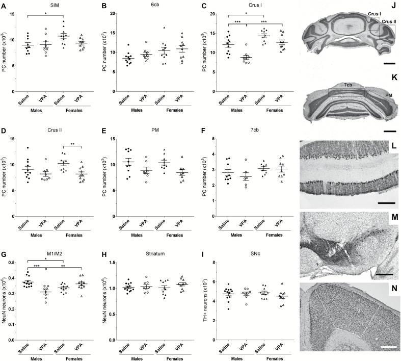
Figure 5.
Loss of neurons in mice prenatally exposed to VPA is sex and region specific. (A–F) Stereological Purkinje cell (PC) count after Nissl staining on coronal section of cerebellum. (J–K) Photos of the different sub-lobules of the lobule VII, scale bars=1 mm. (L) Illustration of the monolayer organization of PC in the cerebellar cortex after DAB-calbindin immunolabeling, scale bars=200 µm. No difference in PC number was found within the hemispheric part of the lobule VI, SIM (A), or in the vermal part of the lobule VI, 6cb (B). Significant loss of PC was found in the sub-lobule Crus I of the lobule VII in VPA males (C), whereas VPA females showed PC cell loss in the sub-lobule Crus II (D) of the lobule VII. (E) No effect of treatment on the number of PC in the hemispheric sub-lobule PM, or (F) in the vermal sub-lobule 7cb of the lobule VII was found. (G) A decrease in the number of NeuN-stained neurons in the M1/M2 motor cortex was found in VPA males (outlined area on N, scale bars=400 µm). (H) There was no effect of treatment on number of NeuN-stained neurons in the striatum. (I) No change was found in the number of neurons expressing tyrosine hydroxylase (TH) in the substantia nigra pars compacta (SNc) after DAB-TH immunolabeling on ventral mesencephalic coronal sections (M scale bars=400 µm). n(A)=(saline males/VPA males/saline females/VPA females)=9/8/10/9; n(B)=11/8/10/10; n(C)=11/8/9/10; n(D)=11/8/9/10; n(E)=10/7/9/8; n(F)=9/6/8/9; n(G)=12/8/11/10; n(H)=12/8/10/9; n(I)=12/8/10/10. Statistical test: 2-way ANOVA with posthoc Fisher’s LSD test. *P<.05, **P<.01, ***P<.001

