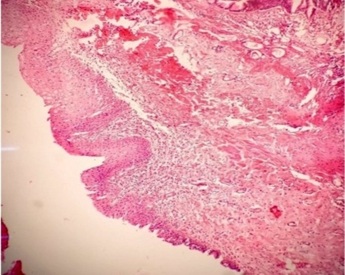
Figure 3.

Photomicrograph at ×10 magnification of biopsy from bladder mucosa under the plaque showing the metaplastic keratinised squamous epithelium on the top left, the transition to urothelium can be seen on the bottom right, along with this there is oedema and inflammatory infiltrates in the submucosa.
