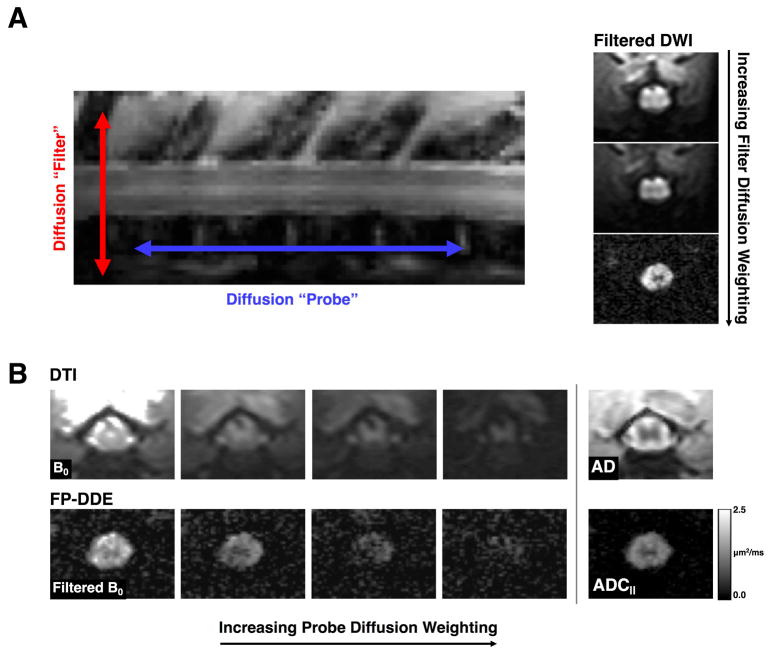
Figure 1. Filter-Probe Diffusion Weighting.
(A) The “filter” and “probe” diffusion weighting directions as used in the FP-DDE collection scheme (left). By increasing the filter diffusion weighting, tissue outside of the spinal cord is attenuated prior to sampling diffusion along the spinal cord (right). (B) Conventional diffusion tensor imaging (DTI; top) employs a single diffusion direction for each measurement with maps of axial diffusivity (AD) reflecting diffusivity along the white matter fibers as well as including all tissue around the spinal cord. The filter-probe double diffusion encoding (FP-DDE; bottom) samples the same diffusion along the spinal cord, but does so after the filter pulse, resulting in a measurement of parallel diffusivity (ADC||). The measures of AD and ADC|| are analogous, but ADC|| maps have removed signals associated with CSF and edema and other tissue, which improves delineation of the spinal cord.

