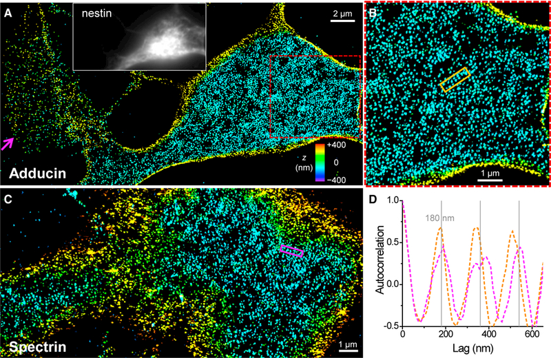
Figure 2. Adducin and Spectrin in Undifferentiated NSCs Are Characterized by Locally Periodic Patterns Commensurate with the Actin Lattices.

(A) 3D-STORM image of immunolabeled adducin at the ventral (bottom) membrane of an undifferentiated NSC. The magenta arrow points to a protruding edge. Inset: immunofluorescence of the NSC marker nestin.
(B) Enlargement of the red box in (A).
(C) 3D-STORM image of immunolabeled βII spectrin (C terminus) at the ventral (bottom) membrane of an undifferentiated NSC.
(D) One-dimensional autocorrelations along the orange and magenta boxes in (B) and (C). Gray grid lines mark multiples of 180 nm. See also Figure S1.
