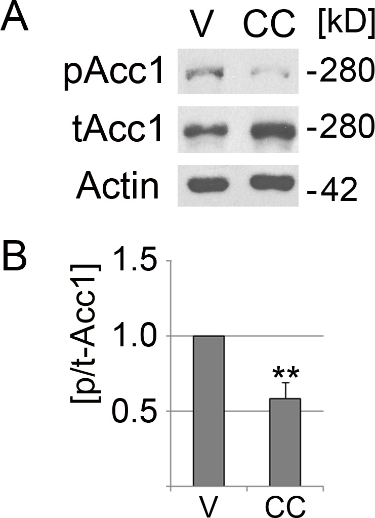
Figure 1. Compound C reduces significantly the phosphorylation of Acc1.

LLC-PK1 cells were incubated with the vehicle DMSO (V) or compound C (CC) for 2 h at 37 °C. (A) Crude cell extracts were analyzed by Western blotting with antibodies against Acc1 phosphorylated on Ser79 (pAcc1) or total Acc1 (tAcc1). Actin was used as loading control. The molecular mass of Acc1 (280 kD) and the position of a 42 kD marker protein are indicated at the right margin. (B) The graph depicts the quantification of ECL signals. For each experiment, the ratio of ECL signals for pAcc1 and tAcc1 (p/t-Acc1) was determined. Data were normalized to the DMSO control (defined as 1.0 on the y-axis). Bars represent the average +SEM of four independent experiments. Student’s t-test demonstrates the significant reduction of the p/t Acc1 ratio upon treatment with compound C; **, p < 0.01.
