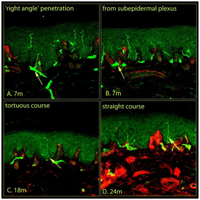
Figure 2.
Morphological features of epidermal nerves within rat plantar skin. Confocal images of glabrous skin biopsies taken from control rats of different age groups with nerves (green) and the basement membrane and dermal structures (red) indicated. The plantar skin samples were removed, post-fixed in the same fixative, cryoprotected in 30% sucrose overnight, and stored at 4°C for batch processing. The stored tissue samples were coded to blind the investigator who performed sectioning, immunohistochemistry, and analysis of the images. Tissues were immersed in Cryo-gel OCT compound and cut into sections 20-μm thick. The sections from all groups were processed simultaneously using standard protocols (See Material and Methods). A series of 1-μm optical sections of the immunostained sections were captured in successive frames with a confocal laser scanning microscope to construct a 3D image (see video, https://youtu.be/IzBwCz-Eu3Q), and stacked into a single image. A, the dermal nerves run along the basement membrane into the dermo-epidermal junction and send fine branches into the epidermis at right angles. B, nerve fibers enter the epidermis from the subepidermal neural plexus located at the dermo-epidermal junction. The nerve endings within the epidermis followed either a tortuous (C) or straight (D) course. Each image captured 634 μm of skin samples.

