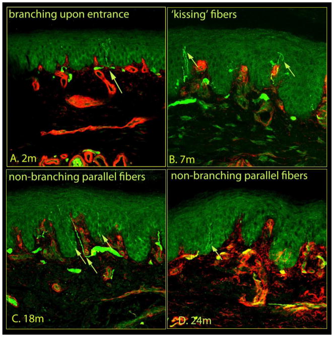
Figure 3.
The morphological features of epidermal nerves within rat plantar skin, in addition to Figure 2. Within the epidermis, occasionally, a nerve fiber branched into two fibers (A) or crossed with another fibers (B) giving a ‘kissing fiber’ configuration. The epidermal nerve fibers were also seen to run in parallel with other fibers following a non-branching (C) and straight (D) course. Each image captured 634 μm of skin samples.

