
Figure 5.
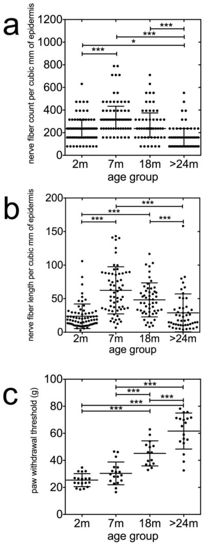
Nonlinear inverted-U shaped relationship between epidermal nerve fiber density and aging. Epidermal nerves were counted as they crossed the basement membrane of the epidermis. For details, see Methods. For measurements of epidermal nerve fiber lengths, images were exported to NIS elements software calibrated to convert pixels to micrometer values, and a polyline was drawn along the length of each identified epidermal nerve fiber and fragment. The density and length of nerve fibers within the epidermis were standardized for section thickness and expressed as numbers per cubic millimeter of epidermis. The median density (a) and total length (b) of the epidermal nerves are significantly increased in samples from the 7-months old (mo) animals and decreased in samples from the >24-mo animals (P<0.05, Krushkal-Walis followed by Dunn’s test). (c) The threshold force required to evoke paw withdrawal threshold (PWT) behaviors were significantly higher in the senescent animals (24–26 mo) compared with the 2-mo (n=18), 7-mo (n=18) and 18-mo (n=15) animals (P<0.0001, ANOVA followed by Newman-Keuls test).