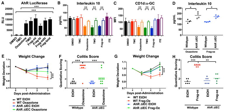
Figure 4. Activation of Aryl Hydrocarbon Receptor by Dietary and Microbial-Derived Oxazole Compounds.
(A) AhR promoter-reporter activity after MODE-K cells conditioned with indicated compounds. ***p < 0.001 (Student’s t test).
(B and C) MODE-K cells were transfected with scrambled (solid fill) or siRNA targeted against AhR (clear fill), conditioned with indicated compounds, and then loaded with α-GC followed by 24.7 iNKT hybridoma co-culture. Quantification of (B) CD1d-restricted IL-10 production. (C) Surface expressed CD1d loaded with α-GC. **p < 0.01, ***p < 0.001 (Student’s t test).
(D) Quantification of IL-10 from overnatant of colon explants 3 days after intra-rectal administration of 1% oxazolone or 1% Frag-oz EtOH (50% v/v) vehicle in WT or animals with AhR-deficiency in epithelial compartment (AhrΔIEC) (n = 4).
(E–H) (E and G) Weight change after intra-rectal administration of (E) 1% oxazolone or EtOH (50% v/v) vehicle or (G) 1% Frag-oz or EtOH (50% v/v) vehicle in WT or animals with AhR-deficiency in epithelial compartment (AhrΔIEC). ***p < 0.001 (Mann-Whitney U-test). (F and H) Quantitative scoring for colitis after intra-rectal administration of oxazolone (F) or frag-oz (H) in EtOH (50% v/v) in WT or animals with AhR-deficiency in epithelial compartment (AhrΔIEC) (n = 5). *p < 0.05; **p < 0.01; ***p < 0.001 (Student’s t test).
See also Figures S4 and S5.

