Fig. 3.

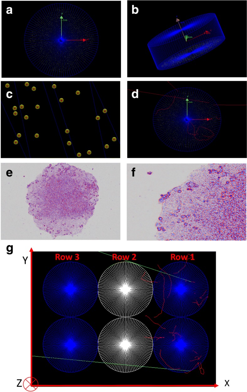
Geometry set-up in the GATE simulations: a, b 5000 non-overlapping spherical cells randomly distributed in a cylindrical well (axial and cross-sectional views); c yellow and red spheres representing the cytoplasm and nucleus respectively; d beta particle tracks (red) in the well; e microspcopy image of a cell culture configuration in a single well; f zoom-in of image in (e); g six adjacent wells in three rows: row 1: active rows with each well containing 288 kBq 90YCl3 in 200 μl cell culture medium, row 2: wells with 200 μl of water, and row 3: with two well only containing 200 μl cell culture medium. The red and green tracks represent emitted beta particles and scattered photons (from the atomic de-excitations), respectively
