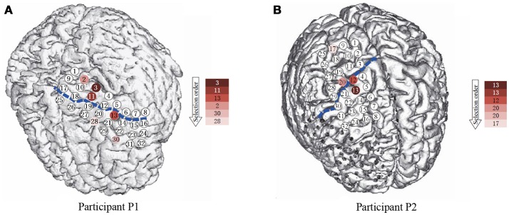
Figure 2.
The spatial position of the subdural ECoG electrodes. (A,B) are the electrode placement for P1 and P2, respectively. The circles are the position of each electrode, the numbers in the circles present the channels. The blue dash lines mark the central sulcus. The color of the electrodes denote the selection priority in greedy feature selection.

