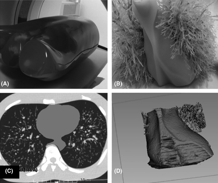
(A) An anthropomorphic chest phantom (Lungman N‐01, Kyoto Kagaku, Co., Ltd., Kyoto, Japan). The anthropomorphic chest phantom was scanned on a multi‐detector CT scanner in order to obtain the volumetric datasets of the original cardiac insert; (B) The original size and the appearance of the cardiac insert; (C) The segmentation process using 3D Slicer software program (The Slicer Community, Harvard).
17 The cardiac insert was segmented to ensure that the modelling process could be performed to produce the heart‐shaped shell; and (D) The virtual 3D model of the original cardiac insert.

