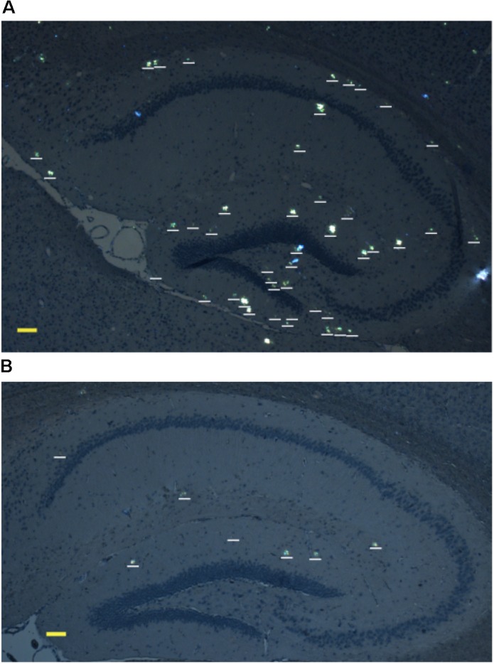
FIGURE 3.

Representative polarized light micrographs of brain sections through the hippocampus for 8-month-old B6C3-Tg(APPswe, PSEN1dE9)85Dbo/j transgenic mice intravenously injected with sterile PS (A) or synthetic isoD7-pS8-Aβ42 peptide (B). Amyloid plaques are highlighted by white bars. Scale bars: (A,B) 100 μm.
