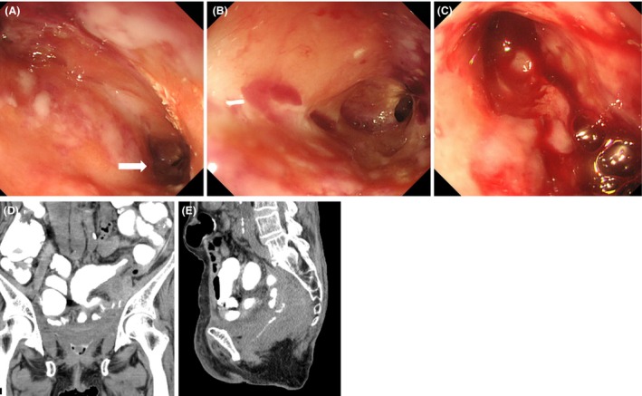
Figure 2.

Videoscope revealing two fistulas in the vagina (A). Close‐up observation of the left fistula (A, arrow) showing the cavity (B). Active bleeding is observed from the right fistula (C). Collection of the contrast medium in the vagina (D) and fistula formations between the ileum and vagina (E) are observed after injecting contrast medium
