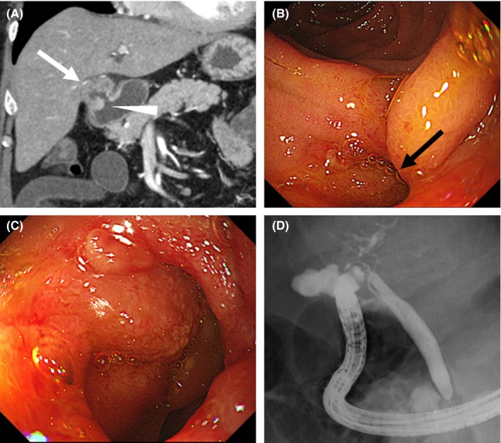
Figure 2.

A, The presence of cholecystoduodenal fistula (arrowhead) was suspected in the computed tomography image at the time of admission. No tumor was found in the gallbladder (white arrow). B,C, Cholecystoduodenal fistula found in the posterior wall of the duodenal bulb (black arrow). D, Gallbladder and bile duct imaged using contrast medium injection from the fistula of the duodenal bulb
